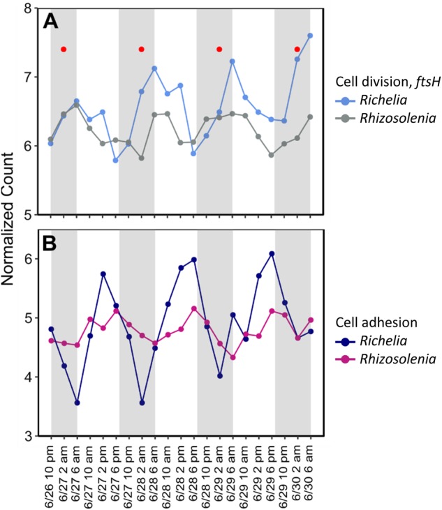
Fig. 7.

Normalized expression patterns of a the putative cell division marker ftsH for both Richelia and Rhizosolenia and b cell adhesion gene forming pili in Richelia and a fasciclin domain contig in Rhizosolenia. All genes/contigs were significantly periodic (RAIN, FDR < 0.05). Red dots in plot A correspond to peak expression timing of silicic acid transporters (Figure S7) in Rhizosolenia. Gray shading highlights dark hours (7 p.m. to 6 a.m.) while light periods are white (6 a.m. to 7 p.m.)
