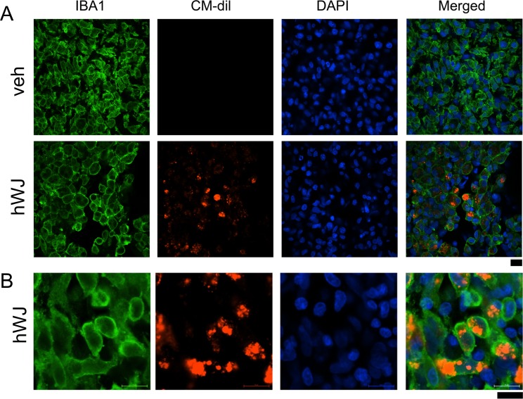
Fig 2.
Transplantation of hWJ-MSCs did not reduce microglial activation in the ischemic core area. hWJ-MSCs were pre-labeled with the fluorescent dye CM-Dil before grafting to stroke rats. Animals were perfused on day 5 after transplantation and stroke surgery. (A) Enhanced IBA1 immunoreactivity (green fluorescence) was found in the ischemic core in animals receiving vehicle or hWJ-MSCs. (A) Red CM-Dil fluorescence (+) cells were confined to the graft sites. No red fluorescence was found in animals receiving vehicle. (B) High-magnification confocal photomicrographs indicated that grafted cells (red fluorescence) were mainly in the microglia. Calibration = 10 μm.
CM-Dil: chloromethyl benzamide 1,1’-dioctadecyl-3,3,3’3’- tetramethylindocarbocyanine perchlorate; hWJ-MSC: Wharton’s jelly-derived mesenchymal stromal cell.

