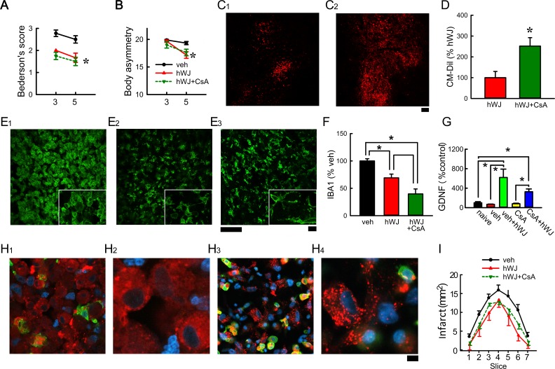
Fig 4.
Treatment with CsA reduced immunorejection of grafted hWJ-MSCs in stroke brain. Transplantation of hWJ-MSCs with CsA (hWJ + CsA) or without CsA (hWJ) significantly reduced (A) Bederson’s neurological score and (B) body asymmetry. Animals were perfused on day 5 after the MCAo. (C) CM-Dil (red fluorescence) was found at the graft sites (C1: hWJ versus C2: hWJ + CsA). (D) A significant increase in CM-Dil fluorescence at the graft site was found in animals receiving CsA at the level of anterior commissure. Compared with vehicle (E1), hWJ + CsA (E3) and hWJ (E2) significantly attenuated IBA1 immunoreactivity in the peri-lesioned cortex (F). High-magnification images (insets) demonstrate ameboid microglial cells in the peri-lesioned cortex in animals receiving vehicle (E1). Ramified microglia were found in the peri-lesioned area in animals receiving hWJ (E2) or hWJ with CsA (E3). (H) CM-Dil (+) cells were found in the lesioned core. Higher magnification confocal photomicrographs indicated that some CM-Dil (+) cells were not contained by the microglia in animals receiving CsA (H2, H4). (I) Brain infarction was examined on day 4 by MRI. The area of infarction in brain slices was quantified every 2 mm from the rostral end. hWJ or hWJ + CsA significantly reduced brain infarction. Calibration C1-2:100 µm; E1–3: 50 µm; E1–3 (insert) 10 µm; H1 and H3 = 10 µm; H2 and H4: 3.2 µm. (G) Transplantation of hWJ-MSCs, with or without CsA, significantly increased GDNF mRNA expression in the grafted side (right) cortex, as compared with the no-transplant controls. The expression of GDNF in the contralateral side (left) cortex was not altered by the transplant.
CM-Dil: chloromethyl benzamide 1,1’-dioctadecyl-3,3,3’3’- tetramethylindocarbocyanine perchlorate; CsA: cyclosporine; hWJ-MSC: human Wharton’s jelly-derived mesenchymal stromal cell; MCAo: middle cerebral artery occlusion; MRA: magnetic resonnace imaging.

