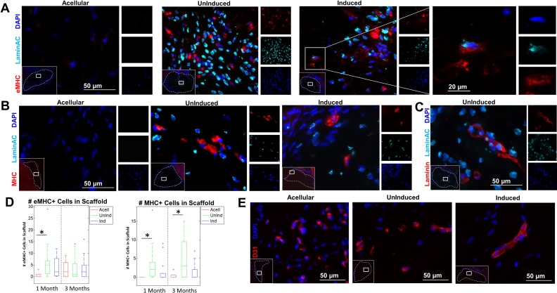
Fig 4.
Immunostaining of embryonic myosin, myosin heavy chain, and LaminAC+ nuclei in the fiber interior. (A) Sections of the excised defect site were stained with embryonic myosin (eMHC; red), human-specific LaminAC (cyan), and DAPI (blue). Inset (left): concentrations of eMHC were found in the center of the implanted fibers. Dotted line denotes fiber boundary. Inset (right): Example of colocalizing eMHC+ and human LaminAC+ cell in fibers with induced ASCs. Images depict fibers at 4 weeks. (B) Sections of the excised defect site were stained with myosin heavy chain (MHC; red), human-specific LaminAC (cyan), and DAPI (blue). Inset: concentrations of MHC were found in the center of the implanted fibers. Dotted line denotes fiber boundary. Images depict fibers at 12 weeks. (C) Sections of the excised defect site were stained with laminin (red), human-specific LaminAC (cyan), and DAPI (blue). Inset: concentrations of laminin+ cells with human nuclei were found in the center of the implanted fibers. Dotted line denotes fiber boundary. Image depicts uninduced fibers at 4 weeks. (D) Quantification of the number of eMHC+ and MHC+ cells inside fibers among the three groups (n=9–15). There were significantly more eMHC+ cells at 1 month in fibers with uninduced ASCs than in acellular fibers. There was no significant difference in eMHC expression among groups at 3 months. There were significantly more MHC+ cells at both 1 month and 3 months in fibers with uninduced ASCs than in acellular fibers. (E) Sections of the excised defect site were stained for mouse CD31 (red) and DAPI (blue). Inset: host vascular infiltration was found in the interior of the implanted fibers in all groups at 3 months. Dotted line denotes fiber boundary.
*p < 0.05.

