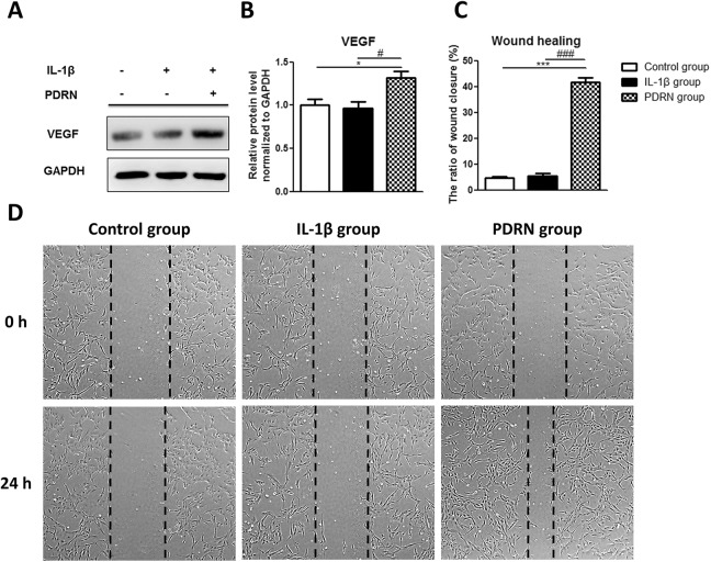
Fig. 3.
Effects of PDRN on protein levels of VEGF and wound healing. (A) Western blot analysis was performed with anti-VEGF and anti-GAPDH (as a control). (B) The relative protein expression in the IL-1β group and PDRN group compared with the control group by western blot. (C) The area of the wound closure was quantified, and the ratio of wound closure was expressed as a percentage of recovered wound compared with the area at 0 h of each group. All results are expressed as mean ± SEM. *p < 0.05, and ***p < 0.001 vs. The control group. #p < 0.05, and ###p < 0.001 vs. The IL-1β group. (D) Representative data of wound healing experiment. The beginning of the experiment is before treatment with PDRN and indicated as 0 h in the figure. After treatment with PDRN for 24 hours is indicated as 24 h in the figure.
GAPDH: glyceraldehyde-3-phosphate dehydrogenase; IL: interleukin; PDRN: polydeoxyribonucleotide; SEM: standard error of the mean; VEGF: vascular endothelial growth factor.

