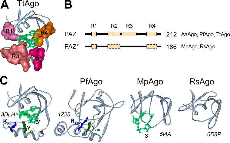
FIG 4.
PAZ domains in pAgo proteins. (A) The structure of the 3′-guide binding pocket in TtAgo (PDB ID: 3DLH). Structural regions R1 to R4 forming the pocket are shown in surface representation. (B) A general scheme of the PAZ and PAZ* domain structures in long pAgos. All PAZ domains can have R1, R2, R3, and R4 regions. PAZ* is defined as variants of PAZ lacking full-length region R3. No regions R1, R2, and/or R4 could be detected in multiple alignments of some PAZ or PAZ* variants (shown by broken boxes), but their actual presence needed to be tested due to their low conservation. The numbers show how many long pAgos have corresponding domain structures. The pAgos with resolved structures having PAZ and PAZ* domains are indicated. (C) The three-dimensional structures of PAZ (TtAgo and PfAgo) and PAZ* (MpAgo and RsAgo) variants in crystallized pAgos and their binary complexes with DNA (for TtAgo) or RNA (for MpAgo) guide molecules. Tyrosine and lysine/arginine residues from R3 in TtAgo and PfAgo, probably involved in guide binding, are indicated. PDB IDs are shown next to each structure.

