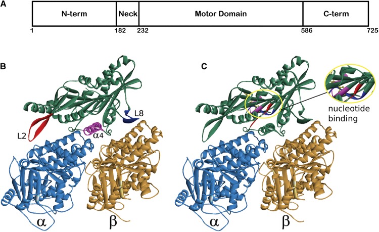
Figure 1. Typical domain layout and motor domain structure of a Kinesin-13.
(A) The domain layout of a typical Kinesin-13, numbering is according to the sequence of the human homologue of MCAK/KIF2C. (B,C) Structure of the human Kinesin-13, KIF2C, in complex with an α/β-tubulin heterodimer (PDB: 5MIO) [42]. (B) The major pieces of secondary structure that defines the microtubule-binding interface are highlighted: Loop 2 (red), α4-helix (pink) and Loop 8 (blue). (C) The location of the nucleotide-binding site is shown inside the yellow oval, plus a magnified view of this region. The major nucleotide-binding motifs are highlighted: p-loop (pink), Switch I (blue) and Switch II (red).

