
Figure 2. Structure of a complex of the human Kinesin-13, KIF2A (green), in a 1 : 2 complex with α/β-tubulin (blue and orange) (PDB: 6BBN) [44].
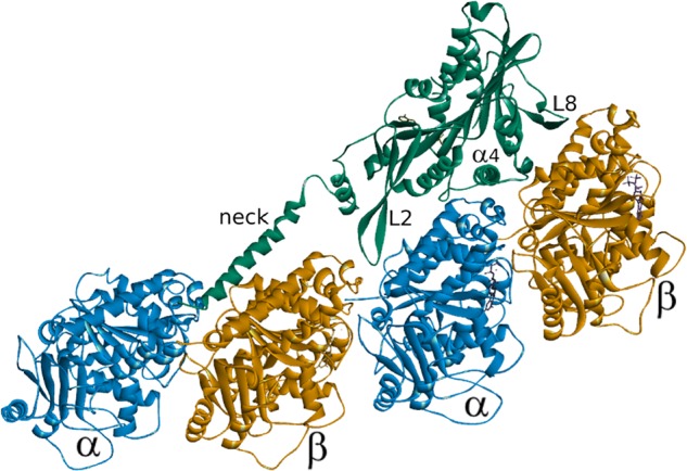
In addition to the major pieces of secondary structure that define the principle microtubule-binding interface, the location of the neck helix is shown interacting with the tubulin dimer on the minus-end side of the tubulin to which the motor domain is bound.