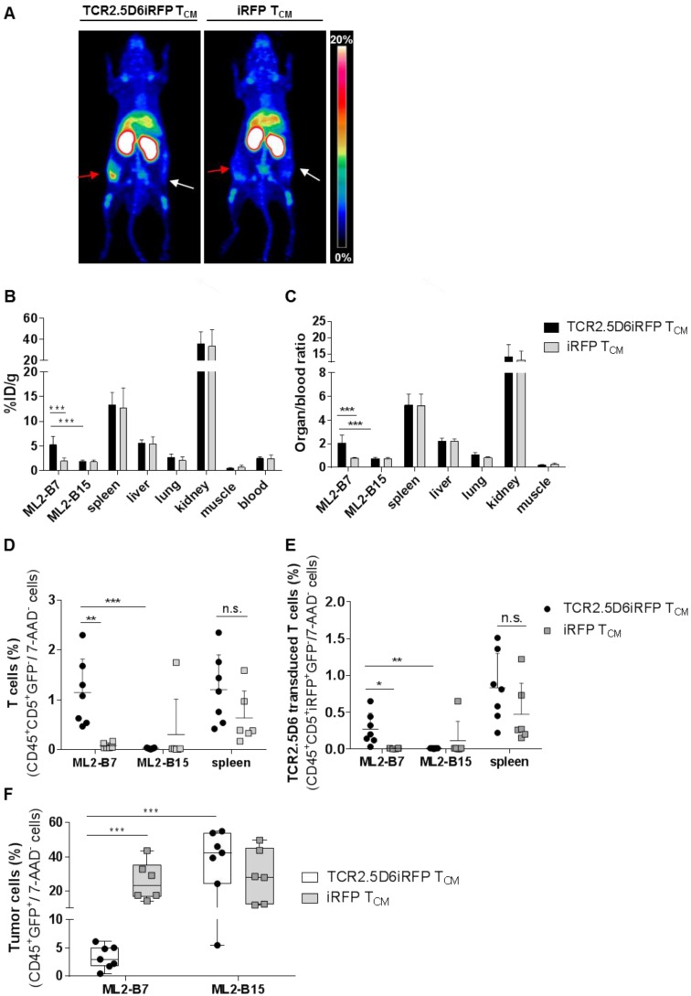
Figure 8.
T-cell tracking by immuno-PET using 89Zr-T3-3A1 (anti-CD7)-F(ab´)2 reveals prominent signals corresponding to accumulation of specific tumor infiltrating lymphocytes at the tumor site. Mice were treated following the imaging protocol depicted in Figure S1A. (A) Representative 3D-MIP PET images of ML2-B7 and ML2-B15 tumor-bearing mice injected with either TCR2.5D6iRFP TCM (n = 7) or iRFP TCM (n = 6). Animals received 89Zr-T3-3A1 (anti-CD7)-F(ab´)2 intravenously and PET imaging was performed 48 h post injection. Red arrows point at ML2-B7 tumor site, and white arrows mark ML2-B15 control tumor site. The scale bar represents 0-20% ID/g. (B) Ex vivo biodistribution analysis of different organs analyzed on the day of imaging. Mean ± SD of %ID/g is shown for different organs from mice receiving either TCR2.5D6iRFP or iRFP TCM. Unpaired t-test: *** p < 0.001. (C) Respective organ-to-blood ratio for TCR2.5D6iRFP or iRFP TCM treated mice. Unpaired t-test: *** p < 0.001. (D) Flow cytometric analysis of T cells detected in the ML2-B7, ML2-B15 control tumor, or the spleen. Mean ± SD of percentage of CD45+CD5+GFP- T cells of 7-AAD- cells is shown for the two groups. Unpaired t-test: ** p < 0.01 and *** p < 0.001. (E) Percentages of TCR2.5D6iRFP transduced T cells of all viable cells analyzed by flow cytometric analysis of respective organs. Mean ± SD is depicted. Unpaired t-test: * p < 0.05 and ** p < 0.01 (F) Percentages of GFP+ tumor cells of all viable cells in ML2-B7 tumors and ML2-B15 control tumors evaluated by flow cytometry. Mean ± SD is shown. Unpaired t-test: *** p < 0.001.

