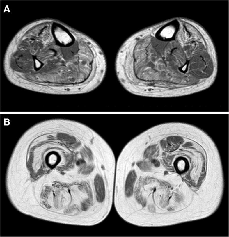
Fig. 1.

T1-weighted muscle MRI at leg (a) and thigh (b) level. In the leg symmetrical fatty changes are more evident in medial and lateral gastrocnemius and, to a lesser degree, in tibialis anterior muscles (a). In the thigh a diffuse fatty substitution is present, with relative sparing of gracilis and rectus femoris muscles (b)
