
Fig. 3.
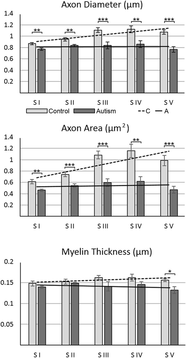
Axon diameter, cross-sectional area, and myelin thickness. In the CC of control subjects, axon diameter and cross-sectional area were the lowest in S 1 but increased significantly in S II–V, and the increment in segments varied in a broad range. In autistic subjects, the average axon diameter and cross-sectional area were significantly less than in the control group, and differences between segments became insignificant. In contrast to variations of axon diameter and cross-sectional area, the average thickness of myelin sheath was comparable in five segments of control subjects. In autistic subjects, significant reduction in myelin thickness was detectable only in S V