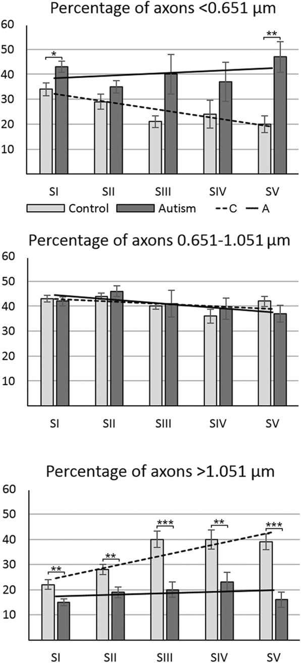
Fig. 4.

Changes of the percentage of small-, medium-, and large-diameter axons in autism. Estimation of the percentage of small- (< 0.651 μm), medium- (> 0.651–1.051 μm), and large-diameter (> 1.051 μm) axons revealed that three features characterized CC connectivity in neurotypical control subjects: significant decrease in the percentage of small-diameter axons in posterior segments, especially in S III–S V; increase in the percentage of large-diameter axons, especially prominent in S III–S V, and a broad heterogeneity of axon size in all segments. The sign of pathological remodeling of interhemispheric connections in CC segments in autistic subjects was stabilization of the percentage of small-, medium-, and large-diameter axons close to approximately 40, 40, and 20%, respectively. The effect of these changes in autistic subjects is the loss of structural diversity of axons typical for a normal brain, resulting in almost flat trend lines for the percentages of small-, medium-, and large-diameter axons in CC segments in autistic subjects
