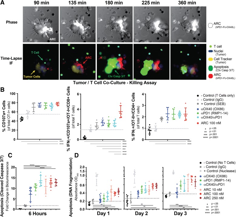
Fig. 6.
PD1-Fc-OX40L Stimulates Tumor Cell Killing In Vitro. a Human NCI-H2023 cells were co-cultured with human T cells stimulated for 2 days with sub-optimal CD3/CD28/IL2. Tumor cells were labeled with a cell tracker (yellow) and nuclear stain (blue), and then cultured with 150 nM of the human ARC, labeled with AF647 (Red). A cleaved caspase-3/7 reagent (green) was added to cells and then the same field was imaged over a time-course. T cells also fluoresced in the green channel and can be differentiated from tumor cells in the phase images (top images) by morphology. Similar to the red channel, the phase images also read out ARC staining (white). Mouse T cells were isolated from C57BL/6 mice (adoptively transferred with OT-I/OT-II cells and vaccinated with Ova/Alum) and co-cultured with B16.F10-ova melanoma cells in the presence or absence of OX40 agonist antibody (OX86), PD-1 blocking antibody (RMP1–14), the combination of those two antibodies or the PD1-Fc-OX40L ARC. b Following 24 h of culture, T cell degranulation was measured by analyzing the proportion of OT-I+/CD8+ T cells expressing CD107a on the cell surface, and IFNγ intracellularly; in both OT-I+/CD107a + cells and also in OT-II+ cells. c Induction of caspase 3/7 cleavage was also evaluated in B16.F10-ova cells in each condition following 6 h of co-culture. d As a late-stage indicator of tumor cell death, a TUNEL assay was performed on days 1, 2 and 3 of co-culture. Results indicate mean ± SD for ≥2 replicates per condition for each of 2 separate experiments

