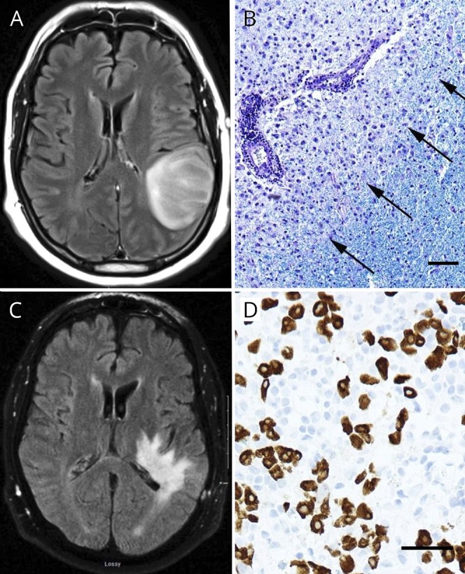
Figure. Images.

(A) Left frontoparietal “mass” (T2 FLAIR). (B) Histopathologic demyelination (Luxol fast blue/periodic acid Schiff), macrophage infiltration, and relative preservation of axons (not shown). (C) Two months later, extensive T2 hyperintensity (T2 FLAIR) believed to represent atypical demyelination. (D) Workup revealed a retroperitoneal mass, with histology positive for germ cell markers SALL-4 (shown) and OCT-3/4 (not shown). Bars = 50 μM.
