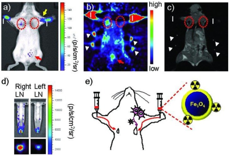
Figure 19.
Triple-modality imaging of radiolabeled nanoparticles: a) optical, b) microPET, and c) MRI of 124I-labeled SPIONs injected into the front paws of a BALB/c mouse bearing a 4T1 tumor implanted on its shoulder. Tumor: yellow arrow; sentinel lymph node: red dotted circle; injection site: “I”; bladder: red arrow; fiduciary markers: white arrow head. d) Ex vivo luminescence (top) and microPET (bottom) images of the dissected lymph nodes. e) Schematic diagram of the tumor metastasis model and injection route of radiolabeled nanoparticles. Adapted with permission from 107, copyright 2010 Wiley-VCH.

