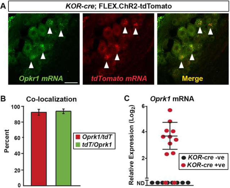
Figure 1. KOR-cre as a tool for targeting KOR-expressing dorsal root ganglia (DRG) neurons.
A.Dual FISH of Oprk1 (green) and tdTomato mRNA (red) shows high co-expression (merge) in DRG neurons. Lumbar DRG neurons were infected with the Cre-dependent virus (AAV.FLEX.ChR2-tdTomato) via IT injection at P40. Arrowheads indicate cells co-expressing Oprk1 and tdTomato mRNA. Scale bar = 25 μm.
B. Quantification of (A). Most tdTomato positive neurons (red) co-expressed Oprk1 mRNA, and most Oprk1 postitive neurons (green) co-expressed tdTomato. n = 3 mice. Data are presented as mean ± SEM.
C. Single-cell RT-PCR of lumbar DRG neurons. KOR-cre mice were infected with an AAV.FLEX.ChR2-tdTomato virus via IT injection at P40. The majority of KOR-cre; AAV.FLEX.ChR2-tdtomato positive neurons (red dots) express detectable levels of Oprk1 mRNA (10 of 12), while none (0 of 22) of the KOR-cre negative neurons (black dots) express detectable levels of Oprk1 mRNA (only 8 cells are shown for clarity; ND, not detected). Data are presented as the -log2 ΔCT expression relative to GAPDH expression within the same cell such that larger numbers represent higher mRNA expression. Dots represent values from individual cells.

