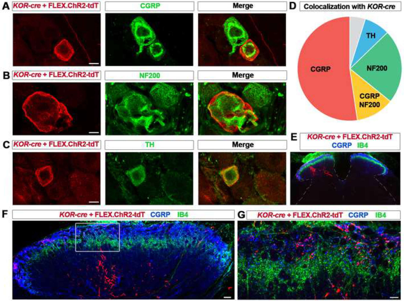
Figure 2. KOR-cre labels distinct neurochemical and anatomical subsets of primary afferents.
A – C. IHC of lumbar DRG neurons from KOR-cre mice labeled using a Cre-dependent virus (AAV.FLEX.ChR2-tdTomato; IT in adult) and co-stained with antibodies to CGRP (A), NF200 (B), and TH (C). Scale bar = 10 μm.
D. Pie chart representing co-localization of immunohistochemical markers with virally-labeled (AAV.FLEX.ChR2-tdTomato, IT in adult) KOR-cre DRG neurons. 52% ± 5% of KOR-cre neurons co-localize with CGRP (red), 12% ± 6% co-localize with both CGRP and NF200 (yellow), 23 ± 6 % co-localize with NF200 (green), and 8% ± 2% co-localize with TH (blue). Only 5% ± 5% of KOR-cre neurons did not co-localize with any of these three markers (gray; n = 3 mice).
E – F. Representative images of lumbar spinal cord section of a KOR-cre mouse following an injection of AAV.FLEX.ChR2-tdTomato into the left sciatic nerve at P40. KOR-cre + FLEX.ChR2-tdt primary afferent terminals can be seen ipsilateral to the injection in the dorsal horn (E) in both the deeper dorsal horn below the IB4 band (F) and in the superficial dorsal horn where they co-localize with CGRP (G).

