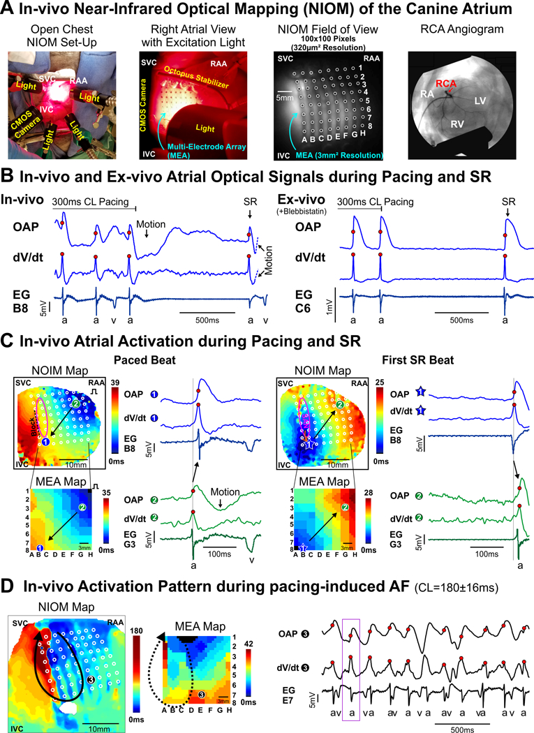
Figure: In-vivo high-resolution near-infrared optical mapping (NIOM) of the canine heart during right atrial appendage (RAA) pacing, sinus rhythm (SR), and atrial fibrillation (AF).

A. Left to right: photograph of in-vivo NIOM camera and lights set-up; close-up photograph of atrial surface with multi-electrode array (MEA) and octopus tissue stabilizer; in-vivo optical field of view from NIOM camera showing MEA (white circles); fluoroscopy angiogram showing the right coronary artery (RCA) where near-infrared voltage-sensitive dye (di-4-ANBDQBS) was delivered. B. In-vivo (Left) and ex-vivo (Right) optical action potentials (OAPs), their derivatives (dV/dt), and unipolar electrograms (EG) from nearest electrode (a- atrial beat; v- ventricular beat) during the end of RAA pacing and the first spontaneous sinus rhythm (SR) beat. C. In-vivo NIOM maps (Top) and simultaneous MEA maps (Bottom) showing activation during RAA pacing (Left) and the first SR beat (Right). Pink oval indicates sinoatrial node (SAN) region. Black arrow represents direction of atrial conduction. Star indicates earliest atrial activation. D. In-vivo NIOM and MEA maps during atrial fibrillation (AF) demonstrate the feasibility of in-vivo NIOM to study arrhythmia mechanisms. Black arrow represents reentry circuit. Purple box indicates mapped time interval. Abbreviations: CL - cycle lengths; IVC and SVC - inferior and superior vena cava; RA - right atrium; RV and LV -right and left ventricles.
