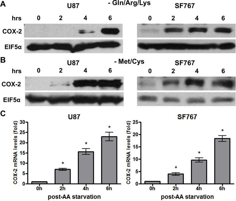
Figure 1.

AA starvation induces COX-2 expression. Indicated cells were cultured in media lacking (A) glutamine/arginine/lysine (Gln/Arg/Lys) or (B/C) methionine/cysteine (Met/Cys). Cells were subjected to starvation conditions for various times and (A/B) probed for COX-2 protein expression on immunoblots or (C) COX-2 mRNA expression by real-time PCR. EIF5α and cyclophilin A served as normalization controls for the immunoblots and real-time PCR assays, respectively. Blots are representative of three independent experiments. Graphs show average value of three independent experiments with error bar representing ± one SEM. * indicate statistically significant (p < 0.05) difference by student’s t-test compared with initial time point.
