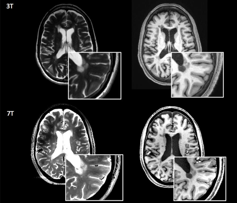
Figure 1.

White matter lesionn detection at 3T and 7T. 3T and 7T axial images obtained for a patient with multiple slcerosis. White matter demyelinating lesions are visualized in greater detail in the 7T T2-weighted (first column) and T1-weighted image (second column). Image resolution: 3T T2-weighted=0.5×0.5×3 mm3,T1-weighted= 0.8×0.8×0.8 mm3; 7T T2-weighted=0.7×0.7×0.7 mm3, T1-weighted=0.7×0.7×0.7 mm3.
