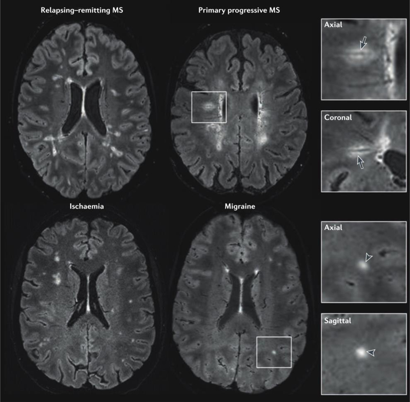
Figure 2.

Perivenous distribution of multiple sclerosis lesions. 3 T FLAIR* (combined T2*-weighted MRI and FLAIR) images from four individuals with a variety of neurological conditions. In the patients with relapsing–remitting or primary progressive multiple sclerosis (MS), a central vessel is visible in most hyperintense lesions (arrows in magnified boxes). On the other hand, a central vein is absent from most of the lesions (arrowheads in magnified boxes) in the patient with migraine and the patient with ischaemic small vessel disease. Reproduced from Sati et al. [21] by permission of Nature Publishing Group (work is licensed under a Creative Commons Attribution 4.0 International License).
