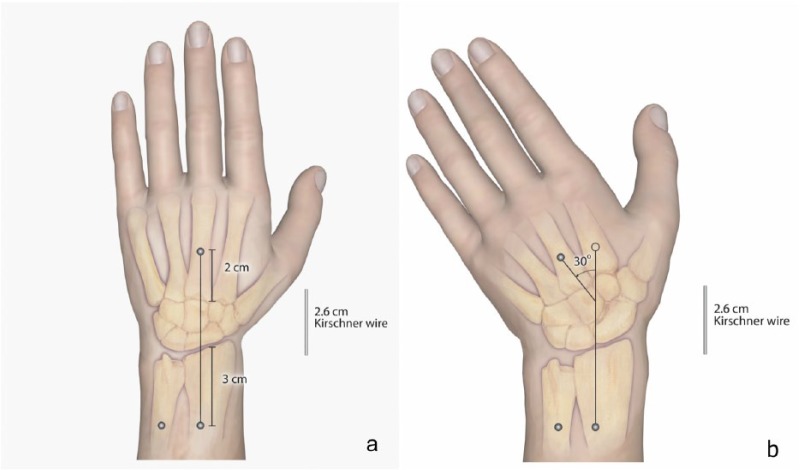
Figure 1.
Illustrations of cadaver positioning.
Note. (a) Each specimen was fixed to a wooden base using headless pins in the radius, ulna, and third metacarpal as shown. (b) Ulnar deviation was achieved by translating the third metacarpal pin to a position in the base set at 30° to the radius shaft. A 2.6-cm Kirschner wire was placed within the field to calibrate digital measurements between all images.

