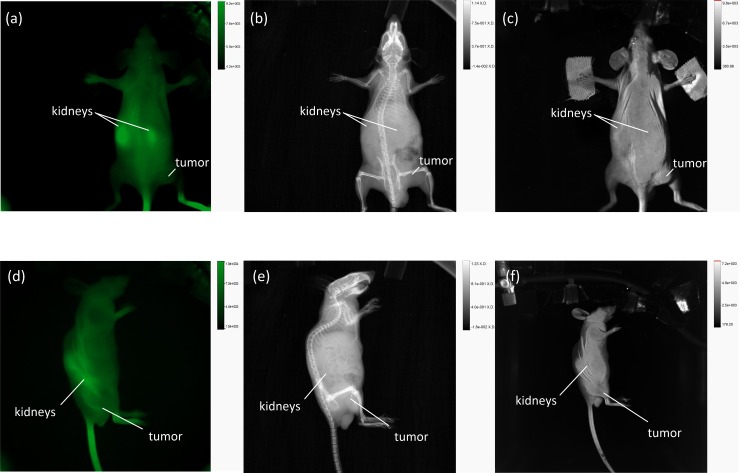
Fig 7. Fluorescence Imaging of AuNP-SIDAG-LUG at 3 h p.i. Using the In Vivo Xtreme System.
(Fig 7A) Fluorescence image at an excitation wavelength of 750 nm to 790 nm. (Fig 7B) X-ray imaging. (Fig 7C) Bright field imaging. (Fig 7D) Fluorescence image at an excitation wavelength of 750 nm to 790 nm from the left side of the mouse. (Fig 7E) X-ray Imaging from the left side of the mouse. (Fig 7D) Bright field imaging from the left side of the mouse.

