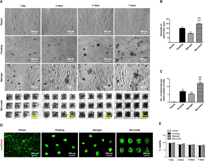
Figure 1.
3D spheroid formation and viability of SGSCs under a priming (spheroid) culture. (A) Floating, Matrigel, and microwell scaffolds induced SGSCs to aggregate and assemble into 3D spheroids in a time-dependent manner. Scale bars = 200 μm. (B) Diameter of spheroids was measured after culture for 3 days, and the values were normalized to the total number of spheroids. The data from five independent experiments were analyzed and presented as a mean ± standard errors of the mean (n = 5). One-way ANOVA, Tukey’s post hoc test. *, compared with plastic; #, compared with floating culture; $, compared to Matrigel. ***P < 0.001, ###P < 0.001, $$$P < 0.001. (C) Spheroid-forming efficacy was determined through measurement of the average number of spheroids per plate after plating the same number of cells. The data from five independent experiments were analyzed and presented as the mean ± standard errors of the mean (n = 5). One-way ANOVA, Tukey’s post hoc test. *, compared with plastic; #, compared with floating culture; $, compared with Matrigel. ***P < 0.001, ###P < 0.001, $$$P < 0.001. (D) A representative LIVE/DEAD fluorescence image of hPECs spheroids after culture for 5 days. (E) Cell viability percentage (viable cell count/total cell count) was measured at 1, 3, 5, and 7 days by the Trypan blue dye exclusion technique. The data from three independent experiments were analyzed and presented as the mean ± standard errors of the mean (n = 3).

