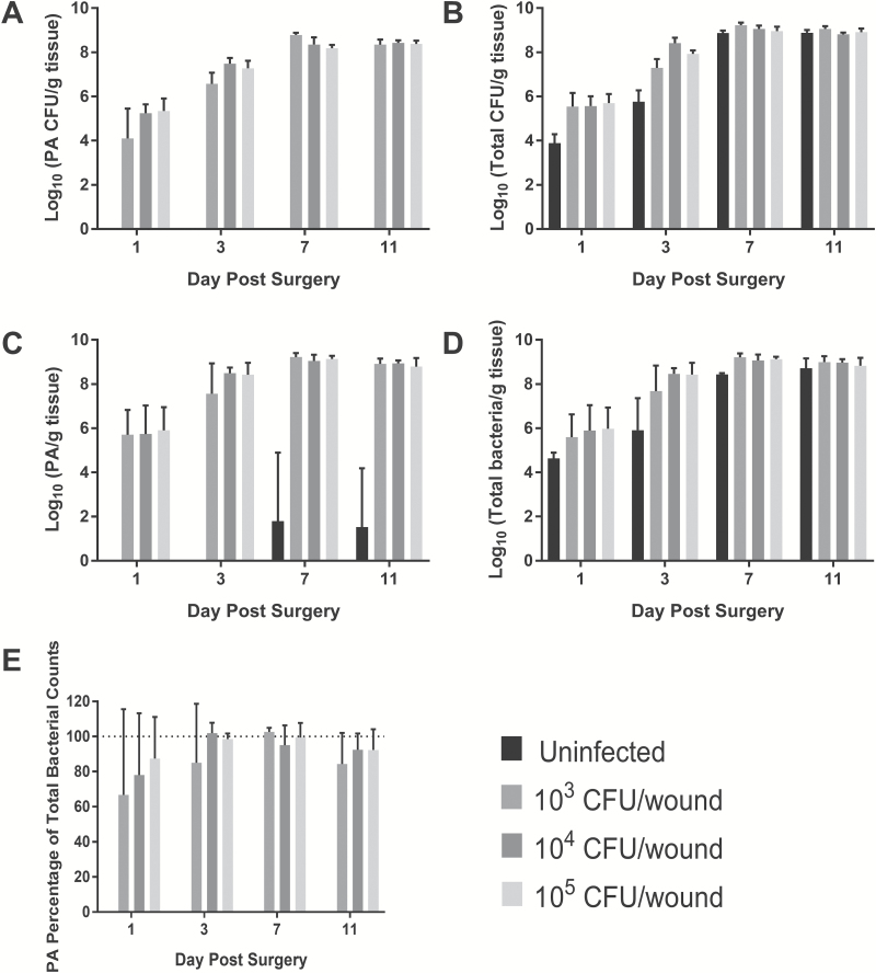
Figure 5.
Bacterial counts recovered from infected partial-thickness burn tissue over 11 days post-burn. P. aeruginosa (A) and total CFU counts (B) recovered as determined by serial dilution and plating on either P. aeruginosa isolation agar or Trypticase soy agar containing 5% sheep's blood, respectively. C. Total P. aeruginosa (live and dead) counts as determined by P. aeruginosa-specific primers. D. Total CFU counts as determined by general gram-negative primers. E. Percentage of total gram-negative bacterial load in the burn tissue that is P. aeruginosa. As time post-infection increased, P. aeruginosa accounted for a greater number of total bacterial cells isolated from wound tissue. P. aeruginosa eventually dominated as the primary wound pathogen. Burns without P. aeruginosa inoculation also became colonized with bacterial cells to the same level as the inoculated groups, but had greater diversity of gram-negative and positive cells. PA, P. aeruginosa.

