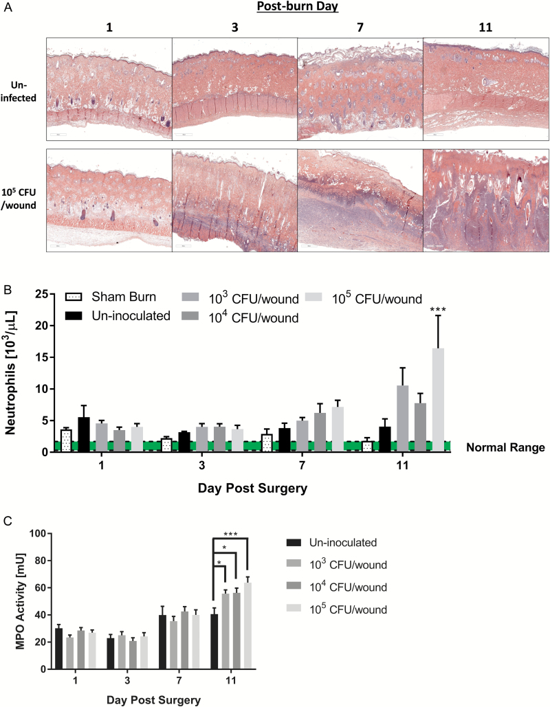
Figure 8.
Histological examination of P. aeruginosa-infected burn tissue. A. Representative hematoxylin–eosin stain tissue sections from un-inoculated and P. aeruginosa (105 CFU/wound) inoculated partial-thickness burn wounds over 11 days following the burn. Compared to the un-inoculated, a large number of inflammatory cells (purple features) infiltrated into the burn eschar in the P. aeruginosa-infected wounds. B. Systemic neutrophil cell counts, prior to euthanasia obtained via cardiac puncture. Neutrophil counts showed increasing trends with P. aeruginosa burn surface inoculation with significant increases detected on post-operative day (POD) 11 between the highest inoculum level and un-inoculated animals (***p < .005, two-way analysis of variance [ANOVA]). C. Myeloperoxidase activity of P. aeruginosa-infected deep partial-thickness burn tissue compared to controls. Significant increases in myeloperoxidase activity were detected on POD 11 in the P. aeruginosa-infected animals, (*p < .05; ***p < .0005, two-way ANOVA).

