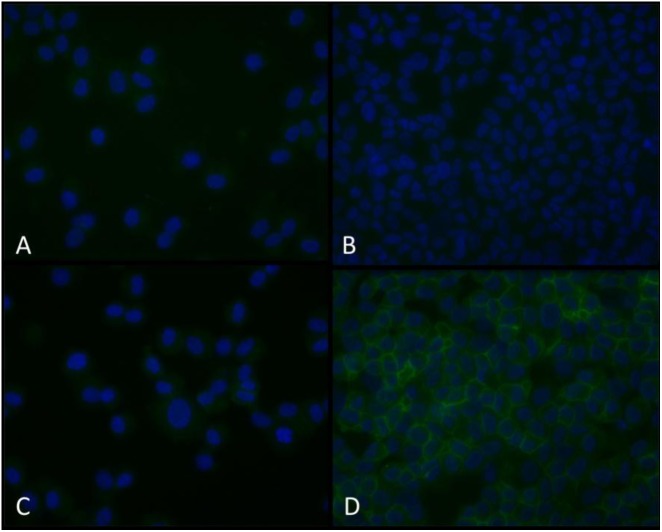
FIGURE 5.
Immunofluorescent NCX1 proteins are increased in TNF-α-conditioned HBSMCs. HBSMCs were grown to near confluence, conditioned without or with 10 ng/ml TNF-α for 24 h and then trypsinized, pelleted on glass slides using a cytocentrifuge, fixed with methanol and probed with a monoclonal antibody to NCX1 as detailed in methods and detected with an Alexa fluor 488 streptavidin-biotin detection system and counter stained with DAPI. (A) Control cells with isotype control antibody. (B) Control cells with NCX1 antibody. (C) TNF-α-conditioned cells with isotype control antibody. (D) TNF-α-conditioned cells with NCX1 antibody. Are the cells presented in each panel from similar donors (Patients undergoing lobectomy or lung transplantation for lung cancer who had no evidence of asthma). Extracellular [Ca2+] from 2 to 0.2 mM.

