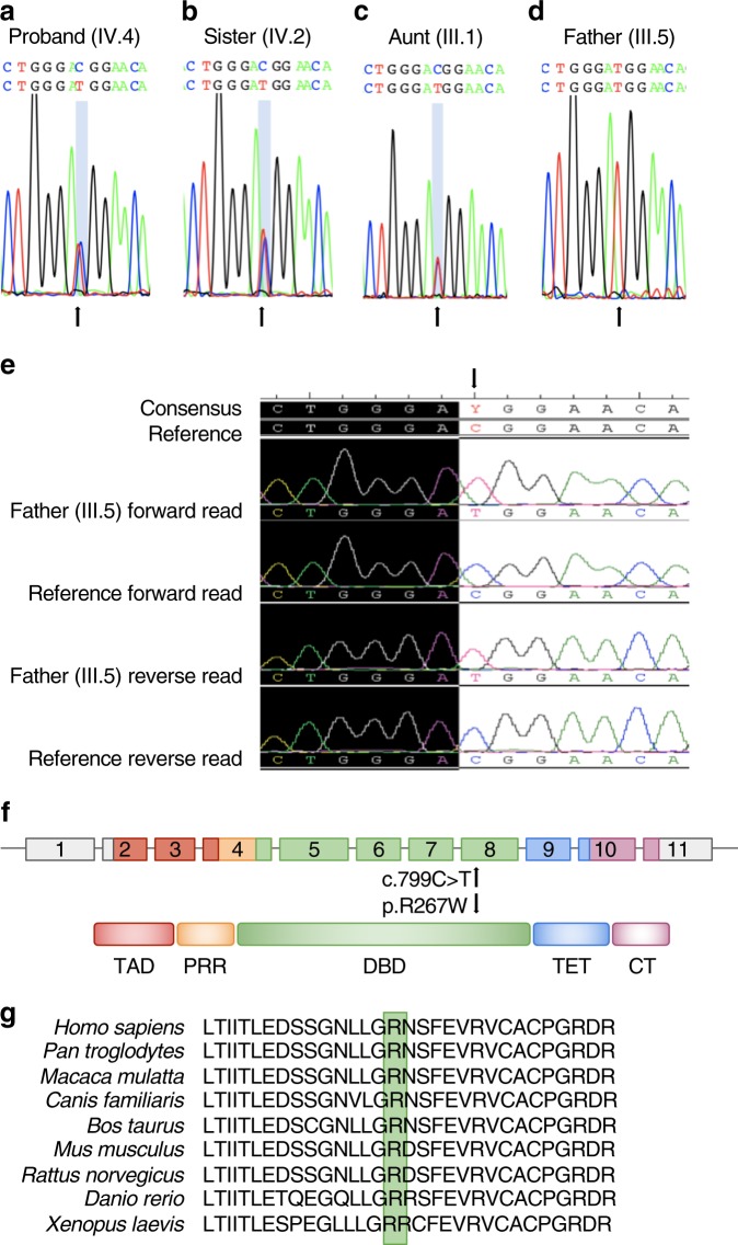
Fig. 3.
TP53 pathogenic variant analysis in the LFS family. a Chromatograms of DNA extracted from saliva showing the heterozygous c.799C > T pathogenic variant (arrow) in the proband (IV.4), b his sister (IV.2), and (c) his aunt (III.1) as well as d the homozygous c.799C > T pathogenic variant in the proband’s father (III.5). e Chromatograms of DNA extracted from peripheral blood showing the homozygous c.799C > T pathogenic variant (arrow) in the proband’s father (III.5) compared to the human genome reference sequence. f Schematic diagram of the human TP53 gene and protein structure. c.799C > T (p.Arg267Trp) is denoted by an arrow. TAD, transactivation domain; PRR, proline-rich region; DBD, DNA-binding domain; TET, tetramerization domain; CT, C-terminal regulatory domain. g p.Arg267Trp is conserved in humans, chimpanzee, Rhesus monkey, dog, cow, mouse, rat, zebrafish, and frog

