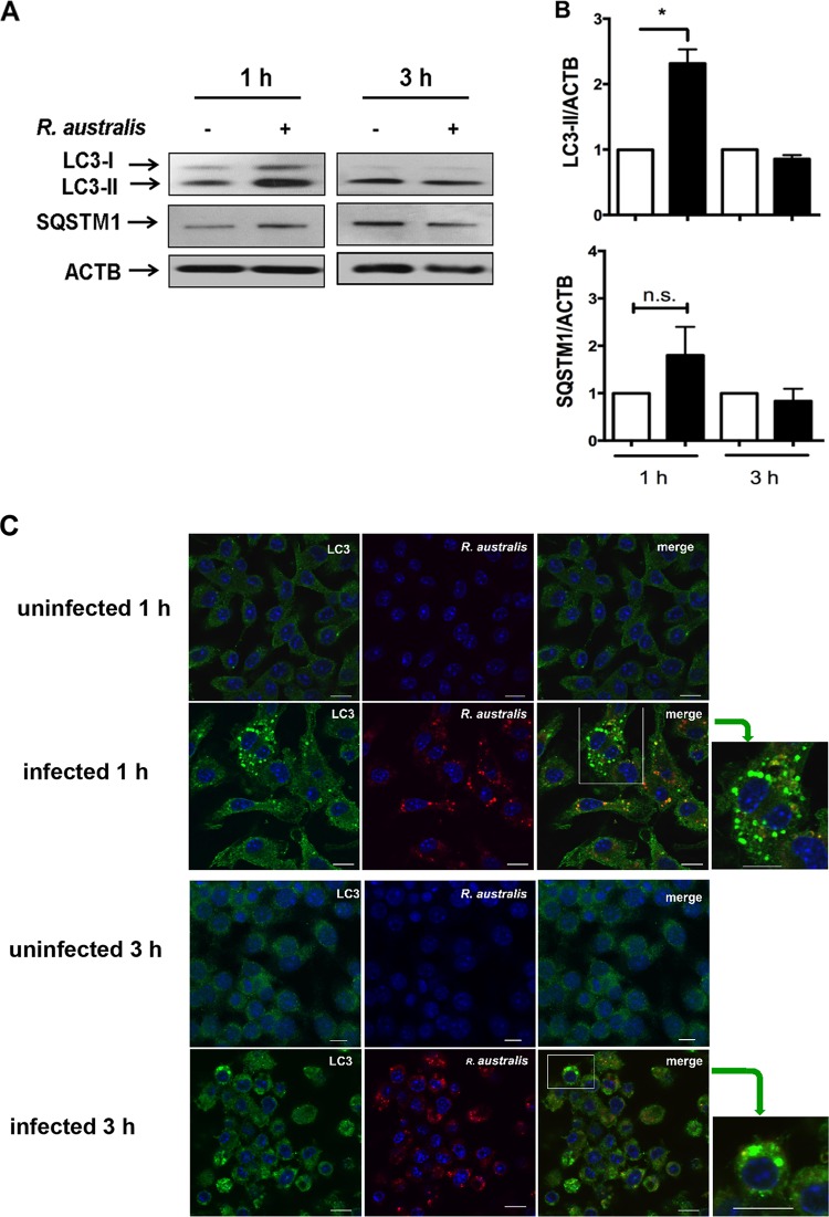
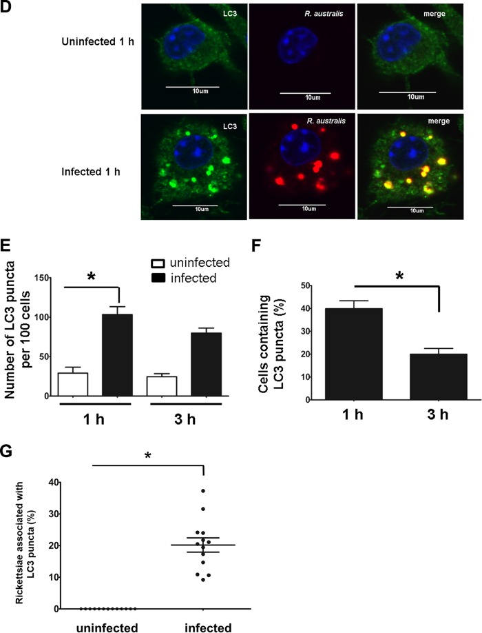
FIG 6.
R. australis infection does not actively induce autophagic flux, although a small portion of cytosolic bacteria was colocalized with LC3 in BMMs from B6 mice. BMMs of WT B6 mice were infected with R. australis at an MOI of 5. Cells were collected at 1 h and 3 h p.i. (A) Cell lysates were immunoblotted with Abs directed against LC3 and p62/SQSTM1. (B) The ratios of LC3-II/ACTB and SQSTM1/ACTB were analyzed by densitometry. (C and D) Representative confocal microscopic images of uninfected and infected BMMs at 1 h and/or 3 h p.i. Bars = 10 μm. Green, LC3 puncta; blue, nuclei (DAPI); red, R. australis. (C) As indicated by the green arrows, the boxes on the right highlight representative cells containing LC3 puncta at a high magnification. (D) The colocalization of R. australis with LC3-positive compartments is depicted in yellow in the cytosol. (E to G) The percentage of cells containing LC3 puncta (E), the number of LC3 puncta per 100 cells (F), and the percentage of rickettsiae colocalized with LC3 (G) were determined. More than 300 cells from 12 randomly selected images were counted at each time point. The data shown are the mean ± standard deviation (SD) from two independent experiments. *, P < 0.05; n.s., not significantly different.


