FIG 2.
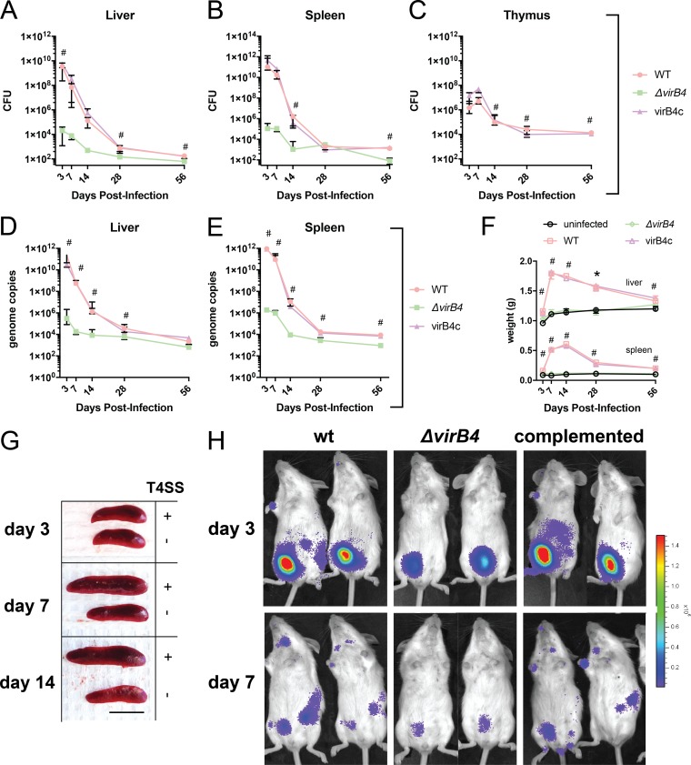
VirB T4SS contributes to pathogenesis. (A to C) Bacterial CFU in liver, spleen, and thymus. (D and E) B. neotomae genome copies in liver and spleen. The data shown are the medians and interquartile ranges for five mice per group. #, significant difference between wild-type and ΔvirB4 bacterial infections using Dunn’s posttest. There were no statistically significant differences for either CFU or genome copies between wild-type and complemented virB4 (virB4c) infections at any time point. (F) Liver and spleen weights during infection. The data shown are medians and interquartile ranges for five mice per group. #, significant difference between both wild-type and virB4c infections and uninfected controls. Liver and spleen weights were not statistically different between ΔvirB4 strain-infected mice and uninfected control mice at any time point. (G) Representative spleens from infections with wild-type (T4SS+) or ΔvirB4 (T4SS−) strains. Significant splenic enlargement was noted 7 and 14 days following wild-type bacterial infection. Scale bar = 1 cm. (H) Small-animal imaging following infection with bacterial strains with a luciferase operon reporter. The color bar is scaled to a minimum and maximum of 2.0 × 10⁴ and 1.5 × 10⁶ p/s/cm2/sr, respectively. Two of three mice per infecting strain with maximal signal are shown. Bacterial inocula in all experiments were 107 per mouse by intraperitoneal injection.

