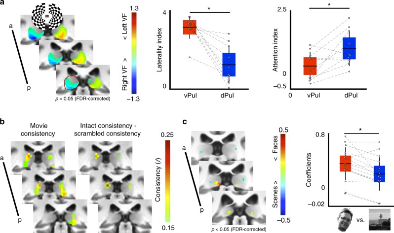
Fig. 1.
Functional distinctions within the pulvinar. a The ventral half of the pulvinar showed greater responses to stimulation of contralateral visual space (left, p < 0.05, FDR-corrected, n = 5). Anatomical extent of the left hemisphere pulvinar in posterior-most slice outlined in dark red. Graphs show the group average (horizontal black line), standard deviation (vertical black lines), and 95% confidence interval (shaded area) for laterality and attention indices. Grey circles illustrate individual subjects. The ventral pulvinar (vPul) showed greater laterality of responses (middle, t(4) = 5.96, p = 0.004, n = 5), while the dorsal pulvinar (dPul) showed greater attentional modulation (right, t(4) = 3.16, p = 0.034, n = 5). b Repeated presentations of intact movie stimuli evoked consistent responses in both the ventral and dorsal pulvinar. Only the dorsal pulvinar showed greater consistency to repeated presentations of intact vs. temporally-scrambled movies (r > 0.15, n = 11). c A posterior medial portion of the ventral pulvinar responded preferentially to face vs. scene stimuli in the voxel-wise contrast (left, p < 0.05, FDR corrected, n = 16) and in the ROI analysis (right, z = 3.10, p = 0.002). Graph conventions same as a

