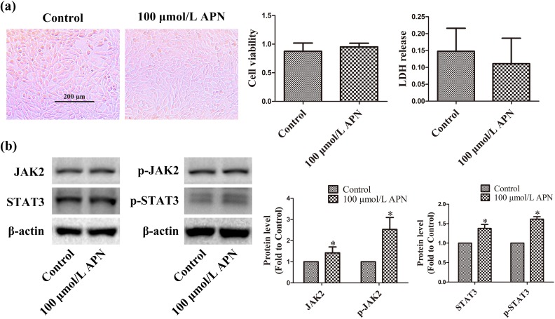
Figure 1.
Effects of APN on HT22 cells. HT22 cells were incubated with 100 µmol/L APN for 4 h. (a) Cell morphology, viability, and LDH release were measured 24 h later. Cells were photographed under an inverted/phase-contrast microscope. Viability and LDH release are expressed as optical density (OD) and absorbance, respectively. (b) Effects of 100 µmol/L APN on the expression and phosphorylation of JAK2 and STAT3 were evaluated using western blot analysis. β-actin was used as a loading control, and the levels of total JAK2, p-JAK2 (Y1007/Y1008), total STAT3, and p-STAT3 (Y705) were normalized to control. n = 6. *P < 0.05, compared with control.

