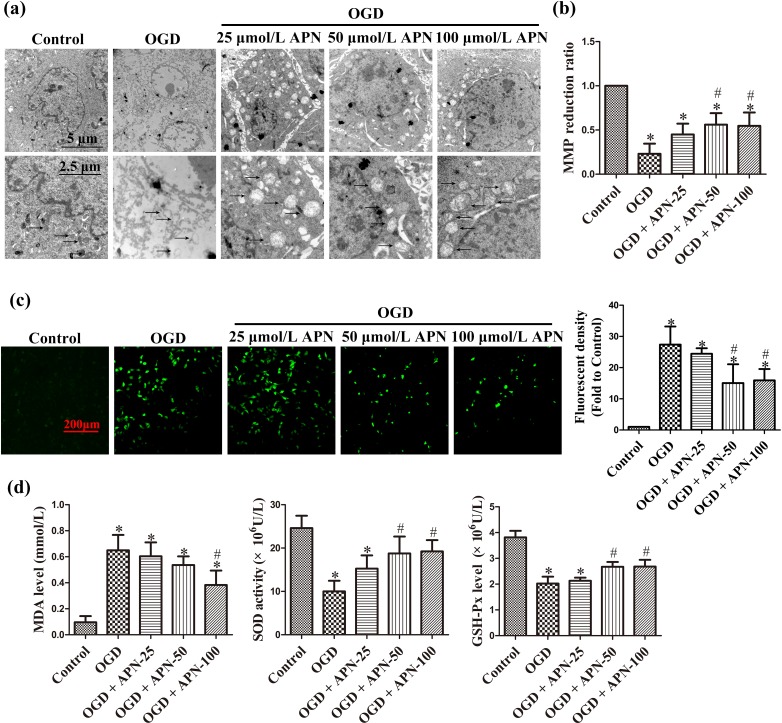
Figure 3.
The protective effect of APN against mitochondrial oxidative injury. HT22 cells were exposed to OGD following treatments of APN (25, 50, or 100 µmol/L) for 4 h. (a) Mitochondrial morphology was visualized by scanning under an electron microscope. Representative mitochondria marked by black arrows. Significant mitochondrial swelling, cristae vanishing, and a reduction in electron-dense substances were observed in OGD compared to control, and these noticeably improved in the APN pretreatment group. (b) MMP was determined by JC-1 staining. The reduction of MMP indicates mitochondrial apoptosis. (c) Oxidative stress level was determined by H2DCF-DA staining. Intracellular ROS were stained with green fluorescence, and the level of ROS was expressed as fluorescent density. (d) Intracellular MDA, SOD activities, and GSH-Px levels were determined using corresponding kits. n = 6. *P < 0.05, compared with control. # P < 0.05, compared with OGD.

