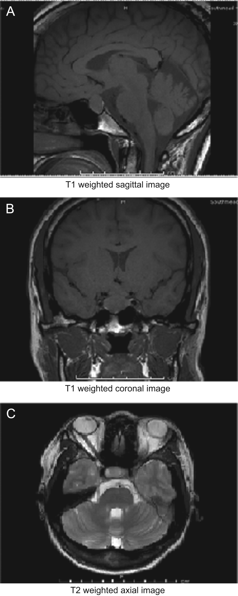
Figure 1.
(A) T1-weighted sagittal image; (B) T1-weighted coronal image; (C) T2-weighted axial image. MRI pituitary showing a mass lesion within the pituitary gland, which is predominantly on the right side. It is 1.5 cm in maximum craniocaudal extent. There is mild contact with the under surface of the optic chiasm, but no clear chiasmatic compression or cavernous sinus invasion.

 This work is licensed under a