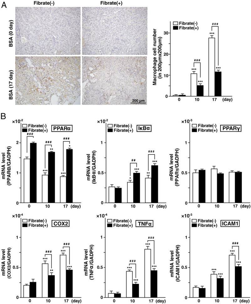
Fig. 6.
Analysis of inflammation. (A) Immunohistochemical analyses of renal tissues. Renal sections were stained for the macrophage marker F4/80. The numbers of macrophages of each group of mice are indicated. (B) mRNAs were obtained from all kidneys in each group of mice. Expression of mRNAs for factors related to the NFκB signaling pathway, including PPARα, PPARγ, IκBα, COX2, ICAM1, and TNFα, were measured with real-time PCR. GAPDH mRNA was used as an internal control. Amounts of mRNA are indicated as target gene copy number/GAPDH copy number. PCR reactions were carried out in triplicate. Values represent means±SD.

