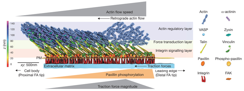
Figure 2.
Nano-scale architecture of the focal adhesion clutch. Focal adhesions (FAs) are organized into 3D ‘nano-domains’ with unique protein compositions and mechanical signatures. The distal tip of the FA facing the leading edgeis where lamellipodial dendritic actin interacts with the FA, and contains an enrichment of phosphorylated paxillin, rapid retrograde flow and high traction forces. The proximal tip of the FA interacts with the actin stress fibre and is enriched with the actin binding proteins α-actinin, zyxin and VASP, and is characterized by slow retrograde flow and low traction forces. Additionally, proteins are stratified in the axis perpendicular to the cell plasma membrane (PM). Paxillin, FAK and the talin head domain are co-localized with integrin cytoplasmic tails near the plasma membrane in the integrin signalling layer. Actin and actin-binding proteins are localized >50 nm above the plasma membrane in the actin regulatory layer. Talin and vinculin reside in the force transduction layer that spans between the integrin signalling and actin regulatory layers. Talin is oriented with the N-terminus near the plasma membrane and the C-terminus ~30 nm higher and extended towards the FA proximal tip. The colour bar shows the vertical distance from the extracellular matrix, whereas the scale bar denotes the distance across the xy plane.

