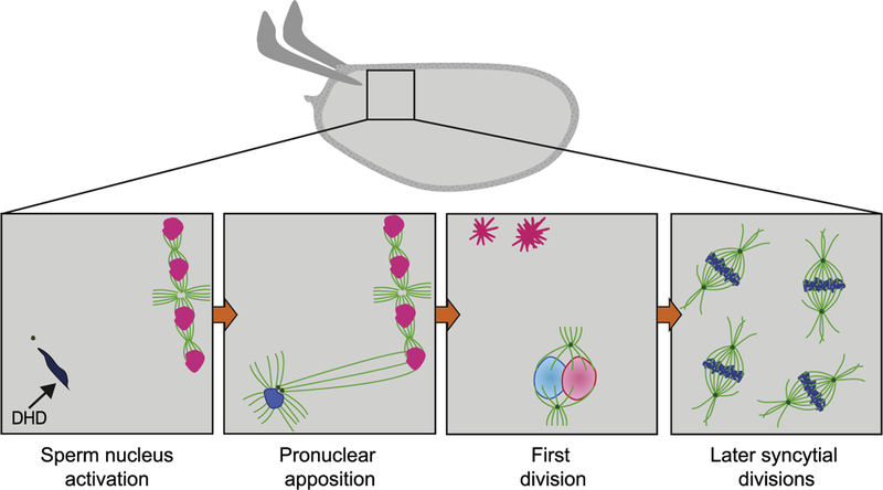
Fig. 2.

Fertilization and the start of embryonic divisions. The activated egg is fertilized once it reaches the uterus. Insets represent progression of cytoplasmic events following fertilization. First, a single sperm enters the egg through the micropyle, and remains at the anterior of the oocyte (dark blue), as female meiosis is completed in the egg and four meiotic products (pink) are produced. The sperm contributes the centrosomes (green) and paternal haploid genomic content (dark blue) to the embryo. Initially the male pronucleus is in a highly-condensed chromatin state due to the presence of protamines. The sperm nuclear membrane breaks down, and protamines are evicted from the sperm DNA and replaced by histones during sperm nucleus activation, in a process that requires the histone chaperone HIRA (not shown) and the maternally provided thioredoxin DHD (shown). Afterwards, the paternal centrioles and MTOCs (green dots) initiate the formation of a mitotic aster (bright green), which increases in size until it reaches the female meiotic product in the closest proximity. The female pronucleus is then pulled towards the male pronucleus during pronuclear apposition. The first division follows, in which there is no intermixing of the maternal chromosomes (light pink) and paternal chromosomes (light blue) until telophase. The remaining female meiotic products undergo chromosome condensation and remain arrested as polar bodies (dark pink) at the cortex of the egg. Subsequent mitotic divisions occur in a common cytoplasm, or syncytia, starting at the middle of the embryo and then expanding towards the surface with increased nuclear number. The syncytial divisions are driven by maternally provided proteins and mRNAs.
