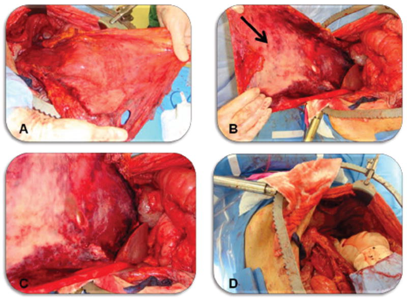
Figure 2.

Radical peritonectomy of the left upper quadrant. A Non-visceral view of the stripped parietal peritoneum from left diaphragm, left paracolic gutter, and left upper abdominal wall stretched to the patients left lower extremity. The ligamentum falciparum and attached pre-peritoneal fat is attached to the medial aspect of the specimen. B Visceral view of stripped peritoneum, arrow marks peritoneal surface disease. C Close view of peritoneal disease burden. D Left upper quadrant status post radical peritonectomy (prior to perfusion).
