
Figure 1.
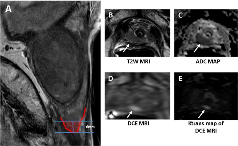
Sagittal T2W MR image shows the distal 6mm apical portion of the prostate corresponding to “distal apical” portion of the prostate (A) in a 72-year old man with a serum PSA of 15.9ng/dL in presence of previous negative prostate biopsies. Axial T2W MRI (A), ADC map of DW MRI (B), raw DCE MRI (C) and Ktrans map derived from DCE MRI (E) demonstrate a right distal apical lesion (arrows). This lesion was sampled with the MRI/US fusion biopsy platform and found to include Gleason 4+4 tumor (90% of core involvement).