
FIGURE 1.
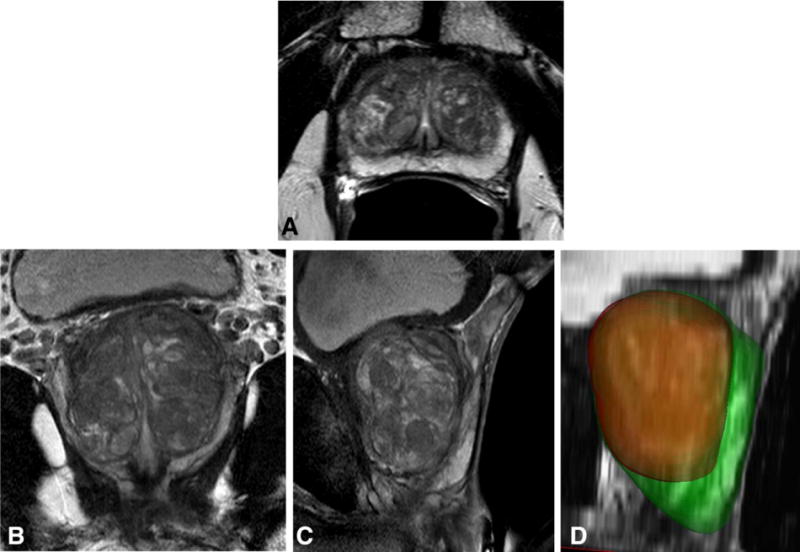
Segmentation results for a 66-year-old patient with PSA level of 6.92 ng/mL, a negative 12-core transrectal ultrasond-guided biopsy, and a negative MR-ultrasound-fusion guided targeted prostate biopsy. Shown are the transverse (A), coronal (B), and sagittal (C) T2-weighted MR images, and the segmentation results of the whole prostate and the central gland (D) in green and red colors, respectively. Based on the segmentation results, the central gland and the peripheral zone are measured 42 and 27 cc in volume, respectively.