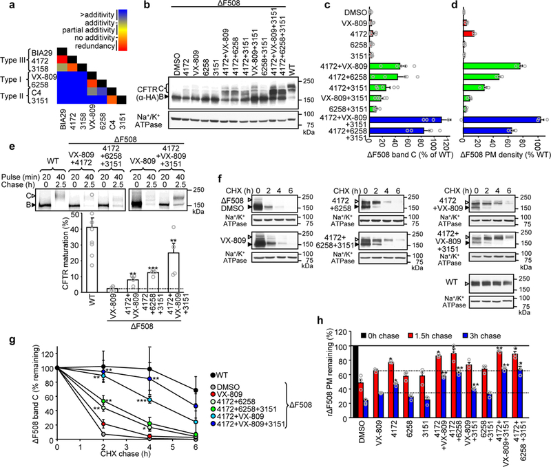
Figure 3.

Structure-guided combination of corrector compounds restores ΔF508-CFTR biogenesis and stability. (a) Combinatorial profiling of compound pair effect on ΔF508-CFTR PM density in comparison to their theoretical additivity (n = 3). The primary PM density data are shown in Supplementary Fig. 3b. (b-d). Effect of indicated single correctors or corrector combinations (4172, 3151– 10 μM; VX-809, 6258 – 3 μM, 24 hours, 37°C) on the expression pattern of ΔF508-CFTR in CFBE41o- determined by quantitative immunoblotting (b) and densitometry (c, n = 5) or measured by PM ELISA (d, n = 3). ΔF508-CFTR values were normalized with CFTR mRNA abundance (Supplementary Fig. 3c) and are expressed as percentage of WT-CFTR control. (e) Determination of ER folding efficiency of WT-CFTR (n = 9) or ΔF508-CFTR in the presence of VX-809 (3 μM, 24 hours, n = 5) or indicated corrector combinations (n = 5 for 4172+VX-809+3151; n = 4 for 4172+VX-809 and 4172+6258+3151) by metabolic pulse chase technique and phosphoimage analysis. The folding efficiency was calculated as the percentage of pulse-labeled, immature core-glycosylated CFTR (B-band, filled arrowhead) conversion into the complex-glycosylated form (C-band, open arrowhead). (f-g) Stability of WT-CFTR or ΔF508-CFTR in CFBE41o- cells upon treatment with VX-809 or compound combinations was determined by quantitative immunoblotting with CHX chase (representative immunoblots of n = 3 independent experiments). The remaining complex-glycosylated (open arrowhead) form was quantified by densitometry and is expressed as percent of the initial amount (panel g, n = 3). (h) The effect of individual compounds or their combinations on the PM stability of low-temperature rescued (48 hours, 26°C) ΔF508-CFTR after 1.5 and 3 hour chase at 37°C (n = 3). Data in c-e and g-h are means ± SEM of the indicated number of independent experiments. *P < 0.05, **P < 0.01, ***P < 0.001 by unpaired two-tailed Student’s t-test in comparison to VX-809 treated samples. The precise P-values are listed in Supplementary Table 4. The uncropped versions of the immunoblots in b and f and of the autoradiographs in e are shown in Supplementary Figure 11.
