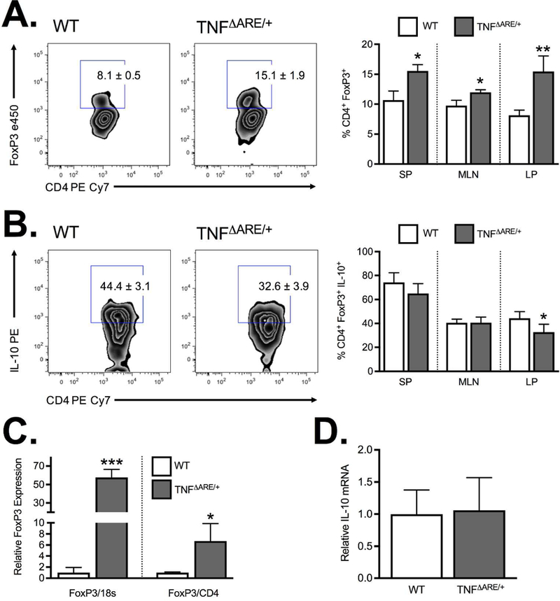
Figure 1. Regulatory T cell expression during ileitis.

(A) Flow cytometric analysis of the frequency of CD4+FoxP3+ from the spleen (SP), mesenteric lymph node (MLN) and ileal lamina propria (LP) demonstrate a significant increase in Treg frequency at peak disease (8–12 weeks) in TNFΔARE/+ mice relative to WT. (B) The increased frequency was offset by a reduction in IL-10 producing CD4+FoxP3+ Tregs in ilea of 8wk TNFΔARE/+ mice relative to WT littermates. (C) Treg accumulation was confirmed by Taqman real-time PCR measurement of the relative FoxP3 mRNA expression in ileal whole tissue with results expressed relative to 18S or CD4. (D) PCR analysis of isolated CD4+CD25+ Tregs from WT and TNFΔARE/+ mice indicated no significant difference in IL-10 mRNA. Results represent mean ± SEM for three mice per group from three independent studies. *P<0.05, **P<0.01, ***P<0.01.
