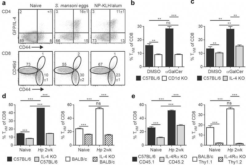
Figure 3.

The expansion of CD8 TVM cells is dependent on direct IL-4 signal. (a) B6 IL-4 reporter mice were immunized with 2500 S. mansoni eggs or 100 µg NP-KLH/alum by injection into the footpad. The draining popliteal LNs were harvested 9 days later and analyzed as described in Fig. 2. (b and c) B6 WT, CD1d KO (b) or IL-4 KO (c) mice were immunized intravenously with 0.5 µg αGalCer. Control mice were treated with solvent alone (PBS containing BSA and DMSO). The spleen was harvested 1 week later and CD8α+ T cells were analyzed as described in Fig. 2. (d) WT or IL-4 KO mice in B6 or BALB/c background either remained uninfected or were infected with Hp. The mesLN cells were harvested 2 weeks later and analyzed as described in Fig. 2. (e) Mixed BM chimeras were generated by reconstituting equal part of irradiated CD45.1+ B6 recipients with WT (CD45.1+) and IL-4Rα KO (CD45.2+) BM, or Thy1.1+ BALB/c recipients with WT (Thy1.1+) and IL-4Rα KO (Thy1.2+) BM. Reconstituted mice either remained uninfected or were infected with Hp. The mesLN cells were harvested 2 weeks later, and WT and IL-4Rα KO CD8 T cells were analyzed by gating on the respective congenic marker. Data are representative of at least two independent experiments with three to five mice per group. Error bars depict the SEM. ns, not significant; **P < 0.01; ***P <0.001 by Student’s t test.
