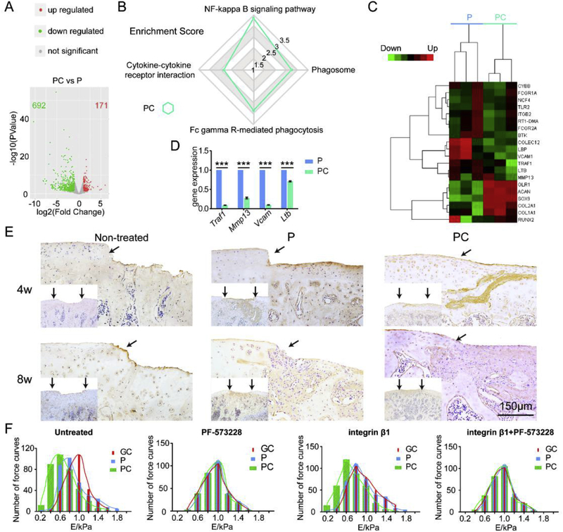
Fig. 7.
Enhanced chondrogenic differentiation of MSCs by PC nanofiber membranes. (A) Volcano plots of gene expression significantly influenced by PC on day 21. (B) KEGG Enrichment Score of PC vs. P on day 21. (C) Heatmap of PC vs. P on day 21. (D) mRNA expressions of Ltb, Traf1, Vcam1 and Mmp13 in both P and PC groups on day 21 using RT-PCR analysis. Mean ± SD; n = 3; *** indicate p < 0.001. (E) Immunohistochemical staining for Il-17 of the engineered cartilage. Scale bar: 150 μm. The black arrows indicate the interface between the repaired tissues and the original cartilage. (F) The histograms of Young’s modulus obtained from AFM characterization of (a) untreated MSCs, (b) PF-573228 blocked MSCs after 6 h, (c) MSCs with AFM cantilever was functionalized by integrin β1, and (d) PF-573228 blocked MSCs after 6 h and with AFM cantilever was functionalized by integrin β1. The cells were cultured on the GC, P and PC nanofibers.

