
Figure 3.
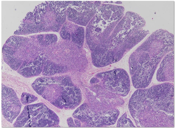
Thymus histologically shown, the outer cortex and the inner medulla. Fibrous septae arising from the capsule penetrate as far deep as the corticomedullary junction creating numerous thymic lobules.
Official websites use .gov
A
.gov website belongs to an official
government organization in the United States.
Secure .gov websites use HTTPS
A lock (
) or https:// means you've safely
connected to the .gov website. Share sensitive
information only on official, secure websites.
Thymus histologically shown, the outer cortex and the inner medulla. Fibrous septae arising from the capsule penetrate as far deep as the corticomedullary junction creating numerous thymic lobules.