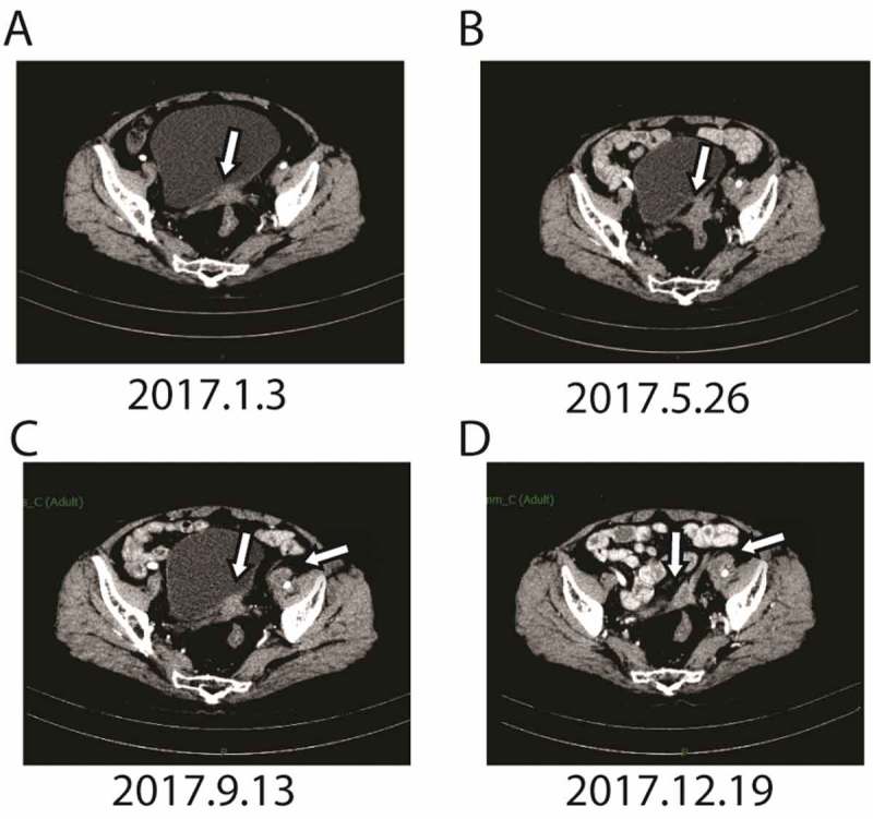
Figure 3.

CT scan before apatinib treatment (the lesions are indicated by arrows). A. after five cycle of apatinib combined with epirubicin, CT scans showed that the metastatic mass became smaller significantly; B after more than four months of Apatinib monotherapy, CT scans showed that the metastatic mass in the vaginal stump was stable. C. after another about four months of Apatinib monotherapy, the mass in the vaginal stump was stable but the lymph node in front of the left external iliac artery increased. D. after another three months, bothe the lesion in the vaginal stump and front of the left external iliac artery were both stable.
