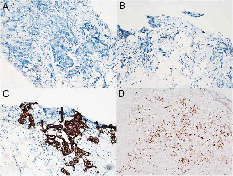
Figure 1.

Immunohistochemical results of the primary invasive left breast carcinoma (original magnification, 200×). The tumor stained negative for both ER (A) and PR (B) but strongly positive for HER-2 (C) and exhibited a Ki-67 proliferation index of 20% (D) .
