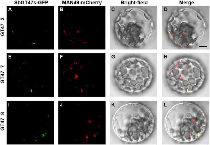
FIGURE 4.
Subcellular localization of SbGT47_2, SbGT47_7 and SbGT47_8. SbGT47s-GFP and MAN49-mCherry (a Golgi marker) were transiently co-expressed in Arabidopsis leaf protoplasts. The localization of SbGT47_2 (A), SbGT47_7 (E) and SbGT47_8 (I) were detected by green fluorescence, while Golgi was marked by MAN49 with red fluorescence (B,F,J). Besides, bright-field images (C,G,K) and merged images (D,H,L) were also acquired for subcellular localization analysis. Bar = 10μm.

