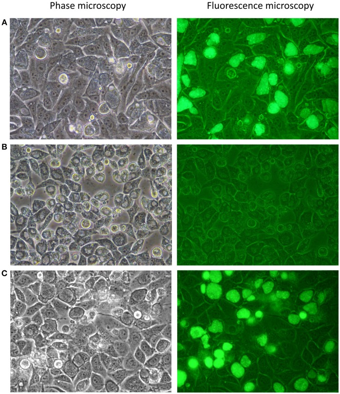
Figure 2.
Photomicrographs of C. trachomatis A2497 strains visualised by confocal and fluorescence microscopy. (A) A2497WT transformed with pSW2::GFP; normal inclusion morphology and green fluorescence are evident, respectively. (B) Plasmid-cured A2497 (A2497P-). Plasmid-free inclusion morphology can be seen by confocal microscopy, typified by a dark border, and pale centre. There is no fluorescence under UV light due to the loss of the GFP gene, which is carried by the plasmid. (C) Restoration of normal inclusion morphology and green fluorescence in A2497P- transformed with the pSW2::GFP plasmid. All strains were grown without selection in the presence of cycloheximide (1 μgml−1) and gentamicin (20 μgml−1).

