Figure 3.
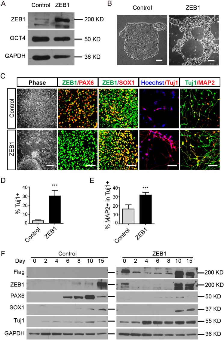
Sustained overexpression of ZEB1 in hESC-induced neural differentiation. A, Western blotting using the indicated antibodies in selected clone #2 of hESCs. GAPDH was the loading control. B, representative phase-contrast images showing different morphologies of H9ESC colonies with (ZEB1) or without (Control) doxycycline in the medium; n = 3. Scale bar: 100 μm. C, images showing the phenotypic alterations of cells at various stages of neural differentiation after overexpression of ZEB1. Typical rosettes in control and extended neurite-like structures from colonies with ZEB1 overexpression were observed. Alterations of PAX6 and SOX1 on day 15, TUJ1 and MAP2 on day 25 were shown as well. Scale bar, 100 μm. D and E, quantification of TUJ1+ cells and MAP2-positive cells. ***, p < 0.001; n = 3. F, Western blotting showed the dynamic expression of ZEB1, PAX6, SOX1, and TUJ1 during early neural differentiation in the control and ZEB1 overexpressing cells. GAPDH was a loading control; n = 3.

